Blog

BBC Crowd Science

(I must remember to update this blog more often!)

I’m typing up a post today because at 20:32 (BST) on the 17th September 2021 I will very briefly be a guest on a BBC radio programme and this may well be one of the biggest outreach opportunities I’ll ever have!

For those of you who know me, you’ll know that I’m very passionate about accessible science and sharing educational resources, so when I was approached to help contribute as a scientist on the Crowd Science episode “How did the human eye evolve?” I literally jumped out of my seat!

I was recruited to talk all things optics (specifically pinhole cameras, refractive index, model eyes etc) although at this point I don’t know what will (or won’t) make it into the show so I’m just very excited to listen in and remind myself of what I said!

The show will be available as a podcast after the show airs at the link below:

https://www.bbc.co.uk/programmes/w3ct1pqy

Check it out!

New Paper!

I’ve recently managed to wade through the notoriously difficult peer-review process well enough to have emerged the other side as a published author!

Please click the link below to read about how we reckon to have found evidence from rTMS for an enhanced role for right V5/MT+ in processing motion presented in contra-lateral *and* ipsi-lateral visual fields

Link to paper *

NewPaper-01.jpg

🌟

Silmo:Paris 2018; quel week-end incroyable

Recently I returned from an incredible weekend at Silmo:Paris where I was invited to give a plenary talk relating to the grant money I won last year. Ultimately, this was such a positive experience that I wanted to share a little bit about it in order to encourage vision scientists to apply for this grant in the future!

First, let’s rewind back to September 2017 when I first heard the incredible news that I was the successful applicant for the Silmo Academy Bursary of €10,000 for 2017. (Please click the link below for a summary of my research). Not only was this a huge opportunity for me professionally and for my research in general, but Silmo wanted to fly me out to Paris to accept the award in person(!). Whilst in Paris I got to watch the previous winner of the award give a fabulous talk about how he used the grant and how useful it was, and I was informed that I would be invited back the following year to give an equivalent talk. Needless to say that after having such a great time, I was very excited to start my research and be allowed the opportunity to return and talk about what I’d done!

Link to Silmo ‘Winners’ Page

If we fast-forward now to September 2018, I attended Silmo D’Or Gala on the Saturday (such a beautiful event, and well done to all the award winners!) and then prepared to give my talk last thing on the Sunday at the Silmo Academy Bursary Symposium. I was fortunate enough to have a truly wonderful audience who not only listened with a keen attentiveness but also asked some great questions at the end. All in all it was one of my most enjoyable presentations to date!

Immediately following my talk the winner of this year’s Silmo Academy Bursary was announced and I’m very pleased to say that it went to an excellent researcher that I know from Anglia Ruskin (Dr Jan Skerswetat) for his research project investigating binocular rivalry and interocular grouping in Autistic Spectrum Disorder.

This means that if you’re at Silmo:Paris 2019 you’ll have the opportunity to hear Jan speak about the work that he does over the coming year; certainly not to be missed.

If you have a research idea and you’re interested in applying for this €10,000 grant, please check back here soon for a link sending you to the Silmo web page full of guidance of how to apply for future rounds. I can say that I recommend it 100%!

Applications for 2019 now closed *~*~

Thanks again to Silmo for the opportunity, for supporting my research, and for hosting such a wonderful event. À bientôt!


What do I research?

Hi!

In honour of my first article being published in print (instead of just early access), I’ve written this little post is to help explain my research to anyone out there who is just interested in what I do! My intention is to take my first first-author paper (found here) and talk through it in broad, understandable terms. In the future I’ll do this for all my papers but for now this is the only one that’s published and worthy of such efforts.

A Direct Demonstration of Functional Differences between Subdivisions of Human V5/MT+

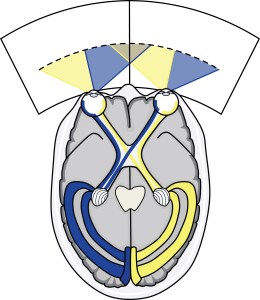
Starting with the title, this is a very informative sentence providing you’re quite well-versed in the field of neuroimaging, motion perception, and neurostimulation. If not, then it probably reads a little strange(!). In essence it’s describing that we’ve managed to determine functional differences between two areas in the brain characterised by their preference for contributing to our perception of moving scenes. These areas (MT/TO-1 and MST/TO-2) are contained within a little region of the brain called human V5/MT+ (1). We have two of these regions, one in the right hemisphere and one in the left, and they can be found in the ascending limb of the inferior temporal sulcus (fold), or roughly speaking, a couple of centimetres behind and above your ears (2).

figure_3

Now, in everyday life, if we had two whisks for example, we might consider giving one away because it’s a waste of cupboard space to have two items that do the same thing, and the same principle applies to brain areas. It would be a waste of precious cortical space to have several visual areas processing the same aspects of the visual world, so if it seems that we have two visual areas that process moving scenes, then it’s a safe assumption that they must be analysing slightly different aspects of the movement. This means we can assume a functional difference without even investigating it, but then proving it is the tricky part…

Now, in order to determine what neurons are processing, we can use neuroimaging techniques such as functional magnetic resonance imaging (fMRI). This works by measuring changes in the level of oxygenated blood in the brain under the assumption that much like muscles, if exerted, more energy needs to be sent to help them to work. If a population of neurons are being used in a particular task then, the heart will need to send more oxygenated blood to replenish the exertion. Previous studies have used this technique to great success and found tentative evidence that both MT/TO-1 and MST/TO-2 respond to different directional stimulation e.g. upward/downward (translational) motion, expanding/contracting (radial) motion, and clockwise/anti-clockwise (rotational) motion, but that perhaps MST/TO-2 shows more preference for the more ‘complex’ radial and rotational stimuli (3-4).

“Why is this interesting?”, I hear you ask…

Well! Typically the higher up the visual processing pathway (usually the more anterior, towards the front of the brain but not always), the more stringent the criteria need to be to drive the neurons. So if MST/TO-2 prefers more complex stimuli it must be higher up the motion processing pathway and that means it’s likely receiving input from MT/TO-1. Cool, right?!

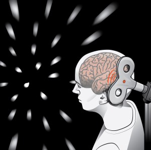
So although using fMRI is the gold-standard approach and is an incredibly informative method for determining what our brains are doing in vivo (while we’re still alive!), our issue here is that we’re just correlating (that is, inferring a relationship between) the increase in oxygenated blood in MST/TO-2 with the presentation of some complex moving stimuli. In an ideal scientific world we seek causal (determining cause and effect) evidence as opposed to correlational. One way of achieving this is to use a technique called transcranial magnetic stimulation (TMS), which (without going into bags of detail) temporarily disrupts the normal cortical functioning of small areas of the brain underneath the ‘coil’ used to apply a magnetic pulse (5). These pulses are very brief and if localised correctly, for example, if we positioned it over MST/TO-2 in someone, then we can design an experiment to test which tasks the participant becomes poor at when they don’t have appropriate use of the MST/TO-2 region.

cer_cor_strong_1

This approach is exactly the design of our experiment, so our aim was to determine the function of MT/TO-1 and MST/TO-2 by applying accurately localised TMS to these regions of the brain in separate trials whilst participants performed various direction discrimination tasks involving translational, radial, and rotational motion.

Ultimately we found that when we apply TMS to MT/TO-1 participants seemed to struggle with identification of translational (up/down) motion but they remained experts at identifying radial (expanding/contracting) and rotational (clockwise/anti-clockwise) motion. However, when we applied TMS to MST/TO-2, participants struggled with the translational motion task as well as the radial motion task.

This suggests that MST/TO-2 is in fact more specialised for these expanding/contracting stimuli than MT/TO-1 which the literature proposes may be linked to our perception of the world moving around us as we move forwards/backwards through it (6). This not only supports the theory that MST/TO-2 might be hierarchically higher up the visual processing pathway than MT/TO-1, but also as both areas seemed to be involved in the perception of translational motion, perhaps MT/TO-1 feeds this signal into MST/TO-2 for further processing… Only further experiments will reveal whether this is the case or not!

So, next time you’re running down a corridor, you can take special note of how the environment appears to be expanding around you as you move forwards and you can think to yourself “hey, my MST/TO-2 region is working really well right now”.

optic_flow_bothways_sm

References

(1) Amano K, Wandell BA, Dumoulin SO. Visual field maps, population receptive field sizes, and visual field coverage in the human MT+ complex. J Neurophysiol 102: 2704-2718, 2009.

(2) Dumoulin SO, Bittar RG, Kabani NJ, Baker CL, Le Goualher G, Pike GB, Evans AC. A new anatomical landmark for reliable identification of human area V5/MT: a quantitative analysis of sulcal patterning. Cerebral Cortex 10: 454-463, 2000.

(3) Smith AT, Wall MB, Williams AL,Singh KD. Sensitivity to optic flow in human cortical areas MT and MST. Eur J Neurosci 23: 561-569, 2006.

(4) Wall MB, Lingnau A, Ashida H, Smith AT. Selective visual responses to expansion and rotation in the human MT complex revealed by functional magnetic resonance imaging adaptation. Eur J Neurosci 27: 2747-2757, 2008.

(5) Walsh V, Cowey AC. Transcranial magnetic stimulation and cognitive neuroscience. Nature Reviews Neuroscience 1 : 73-80, 2000.

(6) Warren PA, Rushton SK. Optic flow processing for the assessment of object movement during ego movement. Curr Biol 19: 1555-1560, 2009.


100% Brilliant

I recently had the pleasure of presenting a 45 minute seminar titled “The Psychology of Seeing” at a CET event in London (100% Optical) and had a wonderful time. I met old friends, new friends, optometrists, dispensing opticians, contact lens practitioners, journalists, designers… And the best part was that people actually thought my summary was interesting enough to spend their Saturday afternoons listening to me chat about Psychology/ Neuroscience! So to everyone who attended my talk and to the organisers who invited me to speak in the first place, I’d like to say: thank you so much! I hope you enjoyed it.

And I’m sure the education content next year will be phenomenal (as it always is, I even sat in on a few excellent talks this year without getting CET points!) so I highly recommend you keep the dates in your diary: 100% Optical, 27-29 Jan 2018.

Below I’ve drafted a tiny (brief) summary of my talk for those who might be interested:


The Psychology of Seeing

Retinal cells transduce light into neural signals which allow our brain to make sense of the visual world. You can’t have successful vision without this partnership!

In neurotypical individuals, nerve fibres from the retina then project to the brain in a very prescriptive fashion as outlined below:

aopsc16_pathway

This means that a scotoma identified using monocular fields tests can predict loci of damage along the pathway, e.g. damage to optic nerve affects one eye, whilst damage to one lateral geniculate nucleus (LGN) affects one hemifield. (Please see my Learning Materials for a little map of which scotoma might link to which part of the pathway).

However we are not all born with fully-functioning, ready-to-go visual brains. Instead neurons are created with the potential to learn how to process these signals and then our early experience determines how successful they become. A good real-life example of this is patching to prevent amblyopia in children – if the visual signal is not appropriately corrected before the ‘critical period’ ends then the neural processing capabilities will suffer.

This leads on to the idea that experience shapes our perception. Imagine we’re trying to identify this image here:
cube
For this we can use a theory of perception from Gregory, 1997 to explain how we might achieve this goal:

greg

At the bottom is the object we’re trying to identify (reality), which feeds into our idea of what it is (hypothesis generator). However, the input itself isn’t the only contribution to our perceptual decision – we also use experience from what we’ve seen before (objects explored/ conceptual and perceptual knowledge) and rules of what we know must be true. This allows us to perceive the 12 random lines as a cube when in fact it isn’t. (Technically it is a subset of well-placed lines on your monitor but to you, it’s a cube! Neat, right?).

We can also use visual illusions to measure this effect e.g. the Ponzo Illusion:

ponzo

Which of the red lines is bigger? The one at the top, right?

(Wrong, they’re the same size… Sorry!!).

But they seem like different sizes because we’re assuming there’s an element of depth and distance to the oblique black lines. That means that for the red line to dissect the black lines at the top and not the bottom, the top line *must* be bigger and therefore we perceive it as bigger. What’s really great is that even though you know this to be true, you can’t un-see it because your brain is still trying to analyse the scene in the same way.

Clearly then, top-down processing plays a huge part in our perceptual understanding of the world and it may not necessarily be true to say that we are all perceiving the same image.

Paper Accepted in Cerebral Cortex – Strong et al., 2017

I’ve just had my first first-author paper accepted into a great journal called Cerebral Cortex. It’s open access so feel free to go check it out via the link below the abstract:

A Direct Demonstration of Functional Differences between Subdivisions of Human V5/MT+

Samantha L. Strong Edward H. Silson André D. Gouws Antony B. Morland Declan J. McKeefry

Abstract
Two subdivisions of human V5/MT+: one located posteriorly (MT/TO-1) and the other more anteriorly (MST/TO-2) were identified in human participants using functional magnetic resonance imaging on the basis of their representations of the ipsilateral versus contralateral visual field. These subdivisions were then targeted for disruption by the application of repetitive transcranial magnetic stimulation (rTMS). The rTMS was delivered to cortical areas while participants performed direction discrimination tasks involving 3 different types of moving stimuli defined by the translational, radial, or rotational motion of dot patterns. For translational motion, performance was significantly reduced relative to baseline when rTMS was applied to both MT/TO-1 and MST/TO-2. For radial motion, there was a differential effect between MT/TO-1 and MST/TO-2, with only disruption of the latter area affecting performance. The rTMS failed to reveal a complete dissociation between MT/TO-1 and MST/TO-2 in terms of their contribution to the perception of rotational motion. On the basis of these results, MT/TO-1 and MST/TO-2 appear to be functionally distinct subdivisions of hV5/MT+. While both areas appear to be implicated in the processing of translational motion, only the anterior region (MST/TO-2) makes a causal contribution to the perception of radial motion.

Read the full article here